In my experiment, I investigated LAMBDA bacteriophages, which are viruses that normally invade and destroy bacteria. They pray on the bacterium 'Escherichia coli', which they can eigher destroy by multiplying their DNA inside them or using the bacterial DNA as a store fir their DNA, where the virus can remain for several generations.
In real life, the bacterium has a protection of these viruses through restriction enzymes, which can cut the virus DNA and make it ineffective. One enzyme always cuts justi n specific loci of a DNA sequence and are therefore unique and result in a unique pattern of sequential pieces of DNA. These sequences are of the same length for ach identical DNA genome, so that when analyzing the DNA of a specific cell, it will always result in the same patterns of DNA cuttings for cells that are identical.
When restriction enzyes have cut the DNA, humans can research and identify the length and the patern of each individual DNA genome, by using the technique 'DNA gel electrophoresis', which works by electrically separating the DNA pieces by length, through an electrical field, where the DNA is put in, so that the DNA moves towards the positive electrode, while resulting in a pattern, because the larger pieces of DNA are more slowly.
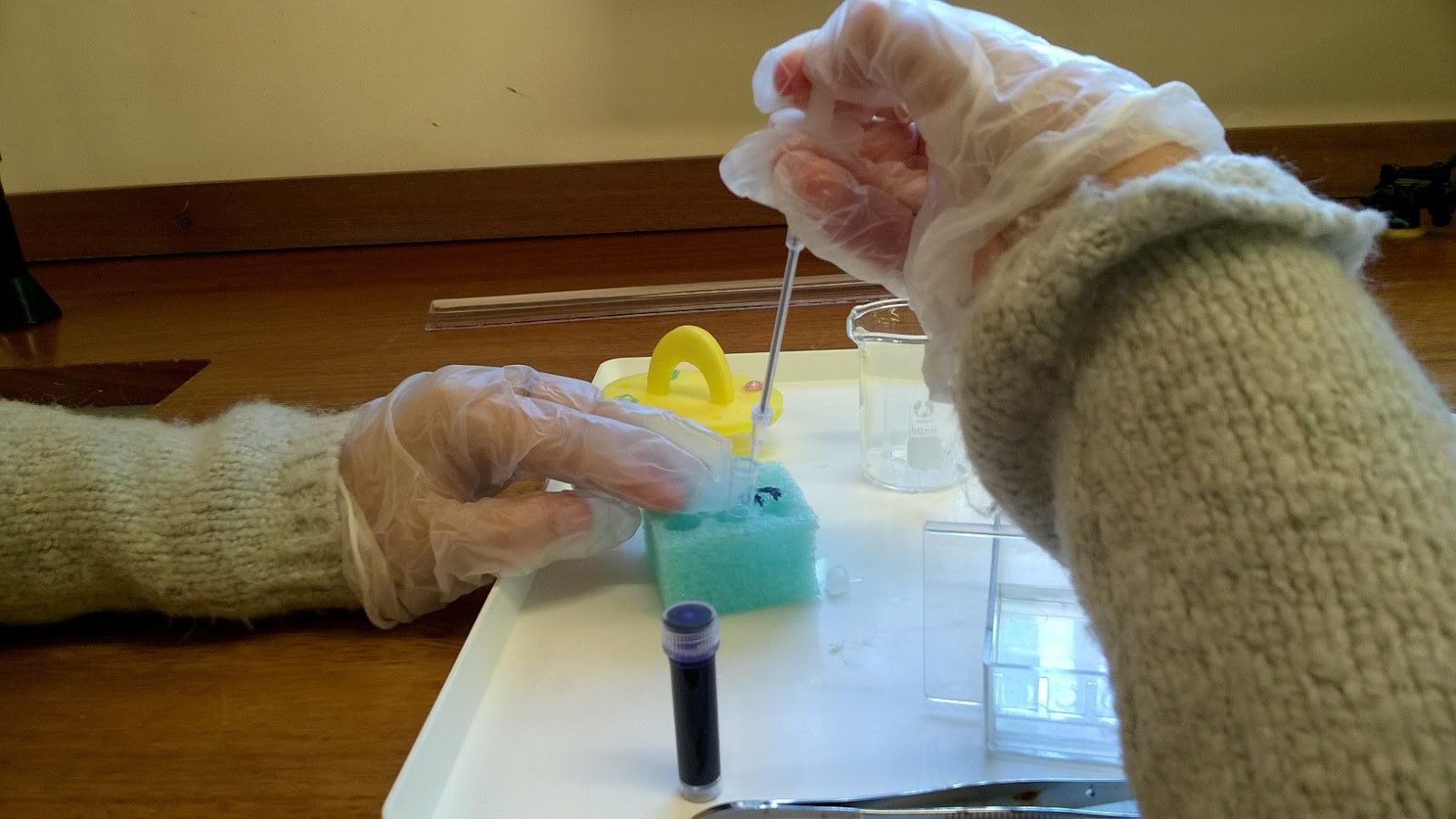
1. Using a microsyringe, I put 20 microlitres of Lambda DNA into 4 coloured tubes, which all contain different kinds of restriction enzymes.
2. Afterwards I placed the tubes in a foam holder and incubated them at a temperature of 37 degrees Celsius. The tubes must stay in the water bath for 45 minutes for the enzyme to work properly.
3. The agrose gel had to be prepared now. Molten agrose(10-12mL) had to be added into an electrophoresis tank, which had to contain a comb, which leaves wells inside the gel, when removing it later.
4. put carbon fibre tissue (electrodes) on each side ofthe tank, so that their is created a positive and negative charge, when later connecting the tissues to the electricity cycle.
5. TBE buffer solution is poured into the tank to make it not to dry.
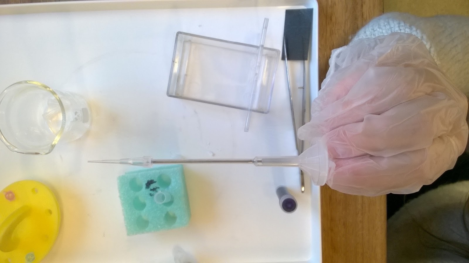
6. Now the different enzyme solutions had to be mixed with dye and loaded into the wells, which are form when removing the comb.
7. After this the gel is connected to a battery, so that the DNA starts running through the gel
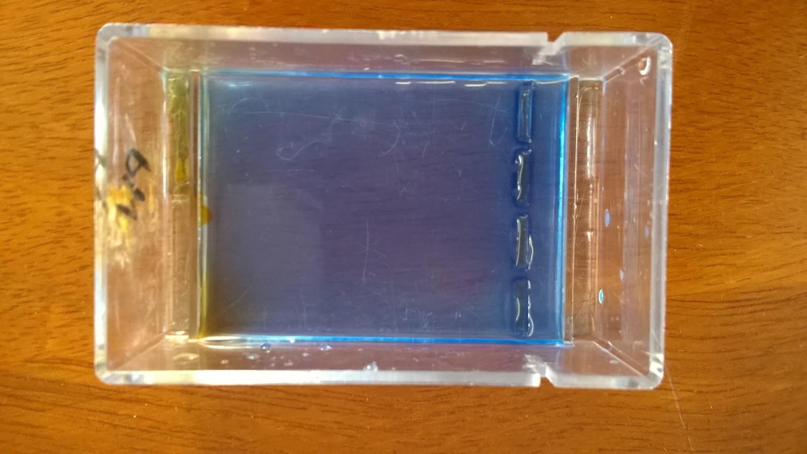
8. Furthermore the buffer can now be poured off and a staining solution has to go on the surface of the gel for 5 min. The stain can now also be removed and washed off additionally with an ethanol solution.
9. In conclusion, the separate parts of the DNA should be visible now.