Donnerstag, 17. November 2016

HIV

How is HIV transmitted?
  • 1)     Contact with blood of HIV infected person through cuts in the skin (also through shared injection needles
  • 2)      Breast milk
  • 3)      Semen and vaginal fluids

History of the disease:
A person gets infected with the HIV virus. 1-2 months after infection, the person gets an infection with fever, headache or rushes. Afterwards, the HIV can stay dormant inside host cells for many years. After around ten years, the immune system is so weak that small infection can already be life-threatening. This stage is called AIDS, when the HIV viruses have killed so many CD4 host cells that there are less than 200 CD4 cells per mm^3.
Basic structure:


Life cycle:
At first, a person is infected either via another person that is infected with HIV.  The HIV virus searches for a host cell in the human blood. This is the CD4 cell, a white blood cell. The HIV virus goes into the the host cell by docking on to a receptor and using endocytosis to enter the cell. There, by reverse transcriptase, it converts its RNA into DNA, which can then enter the nucleus. In the nucleus, the DNA of the virus inserts itself into the cells DNA and thus is transcribed and translated. Thus, proteins are produced for new HIV in the cell. The newly created cells, together with HIV RNA exit the cell by budding of and form new HIV cells.

The effects of HIV on the immune system:
The HIV virus kills the white blood cells, called CD4 cells, which help to protect the body from diseases. Through the progressing destruction of these CD4 cells, the immune system becomes weaker and weaker. When infected, the person will develop a small infection after 1-2 months. In the following years, the HIV can be passive and be dormant in the host cell. After 10 years, the immune system is so weak that a small infection can kill the person.

Why can the body not get rid of HIV?
Because the HIV takes a host cell to replicate, because it does not have an own metabolism. It takes the CD4 cell to replicate inside its DNA. This means, that antiviral drugs would have to aim to kill these CD4 cells, to kill HIV, but through this they would destroy the majorly important CD4 cells, which are part of the immunity of the body.

Why does nobody die directly after an infection with HIV?
Because at the beginning, the HIV has to replicate, because there are too little viral cells to be able o cause much damage to the immune system. Thus, as soon as the HIV viruses enter the body, they will hide in the host cell. Throughout the course of the 10 years of infection, the viruses will slowly destroy their host cells, which can then not anymore defend the body from the HIV virus. Thus, after 10yars the body is finally to weak to fight against HIV.

Opportunistic infections which can lead to death in weakened immune systems:
Salmonella, Pneumonia, bronchitis

Cancers:
Invasive cervical cancer, non-hodkin lymphoma, anal-, liver and lung cancer

Other symptoms:
Fever, chills, rush, mouth ulcers, night sweats, swollen lymph nodes, muscle ache, sore throat

Tests:
A laboratory test, whichtests for HIV antibodies and antigens in the blood is conducted. There is a window period in the first 4 weeks, where you cannot detect HIV infection in your blood, but after 6-8 weeks the reliability of the test increases.

Treatments:
In different stages, different antiretroviral therapy(ART) is done. A combination of 2 are taken in each stage (called antiviral regimen). The drugs are also called antiretrovirals (ARV):
For stage 1: Entry inhibitors (before virus enters host)
Stage 2: Fusion inhibitors (stop virus from being docking on to receptor)
Stage 3: non-nucleoside reverse transcriptase inhibitors, nucleoside reverse transcriptase inhibitors (stopping virus from converting into DNA)
Stage 4: Integrase inhibitors (Stopping virus from joining cell DNA)
Stage 5: protease inhibitors (stopping viral proteins from being produced)

Vaccination:
There is no vaccination on the market yet but scientists are working on it.

Prevention:
Getting tested
Know the sex partner’s HIV status
Use condoms
Don’t inject drugs
Take pre-exposure prophylaxis

Social implications of for infected (voluntary or mandatory):
Stigmatization and discrimination of HIV: Many people refer to it in a prejudiced way and have negative attitudes towards people who have HIV. People can be afraid because of fear of contagion. The infected have a low reputation because many people don’t approve of their sex life or habits (drug use). 


Montag, 26. September 2016

Emphysema: Causes, Symptoms and Biological Consequences

The disease causes the air sacs in the lungs, called alveoli, to enlarge. Cell walls are broken down and damaged, resulting in fewer larger damaged air sacs. Thus, the oxygen uptake into the blood and CO2 return is decreased.

Causes:

·         The most important cause is cigarette smoking
·         Air pollution, dust or chemicals
·         Childhood respiratory disorders
·         Low weight

It is caused by an increased level of phagocytes which inflame the wall of alveoli.
The disease can also be congenital (meaning that it is genetically predisposed). People have an alpha1-antitrypsin deficiency.

Symptoms:

·         Shortness of breath
·         Coughing
Minor symptoms:
·         Lung infections
·         Producing mucus
·         Weight loss
·         Fatigue
·         Blue lips and nails
·         Depression
·         Sleep disorder
·         Headache

Biological consequences:

There is an increased level of phagocytes in the alveoli. These normally destroy antigens by engulfing and producing a protein digesting enzyme called elastase, which kills the antigen. An enzyme inhibitor, called alpha 1-antitrypsin normally protects the lung tissue from the elastase by inhibiting the enzyme.
In smokers the number of phagocytes increases, which can therefore inflame the inner wall of the alveoli, because there are not enough inhibitors to inhibit elastase.

Sources:

https://www.google.co.uk/search?q=emphysema&biw=1242&bih=557&source=lnms&tbm=isch&sa=X&ved=0ahUKEwj2qM_g2q3PAhXBvxQKHV_IDi0Q_AUIBigB#imgrc=yQfTRFNQF7dfpM%3A


Lung Cancer: Causes, Symptoms, Biological Consequences

Cancer generally can be defined as out-of-control cell growth.
There are 2 types of tumour, the malignant and the benign tumour. The malignant tumour can spread to other organs of the body, causing secondary tumours to arise. This is more dangerous than a benign tumour which stays at its original location. The process by which a primary tumour is spread in the body to form secondary tumours is called metastasis.

Causes:

Inhalation of carcinogenic substances (x-rays, tobacco, air pollution, chemicals, dust). These are responsible for damaging the DNA. Free radicals form, which want to steal electrons from other body cells and in the process they damage the DNA. Humans can also be born with a predisposition to develop cancer due to a genetic mutation which makes cancer more likely.

Symptoms:

The cancer undermines the lung’s ability to take up oxygen. It tends to block air way paths and tubules. There can also be bleedings. Another sign might be chronic coughing.
  • ·         Coughing intensely
  • ·         Pain in shoulder or chest
  • ·         Changes in coughed up mucus
  • ·         Coughing blood
  • ·         Chronic pneumonia
  • ·         Chronic bronchitis
  • ·         Difficulty breathing
  • ·         Hoarse voice


Biological consequences:

Cancer can occur in 2 situations: either it can spread through the blood by a process called invasion, destroying healthy tissue, or a cell divides and grows and feeds on blood by a process called angiogenesis.

There is small cell lung cancer (SCLC) and non-small cell lung cancer (NSCLC)

Examples of NSCLC:

  • Squamous cell carcinoma:
  • ·         Most common
  • ·         Most common in men
  • ·         Forms in lining of the bronchial tubes

  • Ardenocarcinoma:
  • ·         Most common in non-smokers
  • ·         Most common in women
  • ·         Forms in mucus producing glands in the lungs


Sources:
https://www.google.co.uk/search?q=squamous+cell+carcinoma&espv=2&biw=1242&bih=602&source=lnms&tbm=isch&sa=X&ved=0ahUKEwit_YCJ2K3PAhVFKsAKHVc9BU0Q_AUIBigB#imgrc=MLiJCKzMn8aLuM%3A
https://www.google.co.uk/search?q=Adenocarcinoma&espv=2&biw=1242&bih=602&source=lnms&tbm=isch&sa=X&ved=0ahUKEwizqYyP2K3PAhVqI8AKHR76B6cQ_AUIBigB#imgrc=J2z4VMXs7e_2OM%3A


Sonntag, 25. September 2016

Olaf the oxygen malecule and his journey through the human body

Hello I am Olaf, the oxygen molecule and I want to take you around the body and show you my daily life, supporting the human respiratory system...

You can imagine my cycle around the body like a day at work, starting in the morning when I go to my work place. My state from floating around in the air changes because person x inhales my through his mouth. I am sucked into the trachea then, which is like like walking along a street.

This happens because of the change of pressure in the internal body system and the external pressure. The diaphragm moves downwards pushing the abdomen out and the rib cage moves upwards and outwards at the same time, so that the lungs have more space. Thus, there is an increased volume which decreases the pressure inside the lungs, causing the air to be inspired.

So I travel down the pathway from trachea to the lungs, starting with big tubes called bronchi, which separate into smaller tubes called bronchioles which end in little air sacs, called alveoli.

Arrived at the alveoli, now I have to cross the membranes to get to the blood stream, flowing through a network of little blood vessels called capillaries. This is like a crossing over a pedestrian crossing. I have to float through two cell walls, one of the alveoli and one of the capillary, having each two membranes, thus until arriving in the bloodstream crossing a total of four cell membranes, which is a relatively short way for me, making my day less complicated.

Now, I jump onto a red blood cell which will carry me through the stream, like a personal motor boat. Meanwhile I oxygenate the deoxygenated stream. Special about it is, that it is an artery stream, which I travel along in direction of the heart now, which can be imagined like travelling in the opposite direction of the traffic, because normally arteries lead away from the heart. To jump on the boat and to be bound to a hemoglobin molecule, I have to overcome the wall of the red blood cell, which is like opening the door of a car.

The red blood cell takes me to the heart, which is like a junction, where the direction of traffic changes. Now I am pumped together with the red blood cell into a new artery, which brings me to whatever location where engergy is needed. Arrived at any place in the body, I can mix with my colleagues, a few Mr Water and a particular Mr Glucose. Together, we can produce ATP.

This is basically the important bit of my work and afterwards I am really exhaused and I head of into direction of the outside world. Thus, another red blood cell picks me up via a vein. I have to cross its cell wall again then, binding to a carbohemoglobin that is connected to the cell. On the way back, we cross the juction at the heart again leading to another vein that belongs to the pulmonary system, which my day had started with, the circulation to the cells for energy having been the systematic circulation.

The vein takes me back to the lungs where I have to at first get off the blood cell by exiting through its membrane and then cross the four membranes of capillary and alveoli again, ending in the alveoli as a carbon dioxide compound.

Now, the only step that is left, before my final destination, returning to my home, the air, is to be pumped up by exhalation of person x again. Ventilation is activated by the diaphragm, a muscle which relaxes and pushes upward into a domed shape and the rib cage being pulled in and down. I am moves outside through the bronchioles, bronchi and trachea again, finally floating through the mouth and being released into the atmosphere.

This was my daily routine and the formation of O2 into CO2 in the human respiratory system.

Sources:
http://de.slideshare.net/sarahfree/oxygen-through-the-respiratory-system
https://prezi.com/9uqpc_zz4x1l/oxy-the-oxygen-molecules-journey-through-the-respiratory-system/
http://image.slidesharecdn.com/lecture7resp-141208082139-conversion-gate01/95/respiratory-system-7-638.jpg?cb=1418029376


Samstag, 17. September 2016

COELIAC DISEASE - WHAT IS IT ABOUT?

What celiac disease is:

The lining of the small intestine after the immune system has attacked the gluten
It is an autoimmune disorder. This means that the body attacks its own tissue. This results in inflammation of organs which can be really dangerous. Celiac disease affects the gastrointestinal tract, because the body recognizes gluten, found in food such as barley, rye and wheat as a threat to the body and attacks it. While attacking the gluten, the villi in the small intestine get also damaged through the inflammation and are shortened, which means that they decrease in surface area. This means that they can’t absorb nutrients as efficiently anymore and this results in nutrient deficiencies and so on.

Description of the basic cause and symptoms of celiac disease:

Celiac disease is caused by the body’s own immune system, which reacts and attacks the gluten found in the gut. The disease can be caused partly environmentally and partly by genetics and occurs more often in people who already suffer from another disease. It can develop spontaneously and can be unnoticed by the person who has got it. The villi in the small intestine are damaged and thus nutrients can be absorbed properly anymore. This damage in the intestine leads to symptoms such as
·         weight loss due to malnutrition caused by the poor absorption.
·         diarrhoea and abdominal pain and swelling due to the inflammation.
·         food intolerance
·         inflammation increases the risk of gastrointestinal cancers (e. g. cancer of oesophagus or small intestine)
·         iron deficiency due to lack of red blood cells
·         vitamin deficiencies
·         low bone mineral density
·         visible affects: enamel of teeth, chronic fatigue, joint pain, poor growth, delayed puberty, miscarriage and infertility
·         Neurological symptoms: Migraine, depression, ADHD, epilepsy

Explanation of the effect of celiac disease on intestinal villus and how this leads to its symptoms:

The villus is inflamed and thus shortened and flattened so that nutrients cannot pass through it anymore. This causes the cells to be more sensitive so that cancer can be developed. It also means that nutrients such as vitamins cannot be taken up into the blood, so that vitamin 12 and folate deficiency anaemia can be developed. Iron deficiency also causes a lack of red blood cells, leading to other health problems, connected to oxygen transport.

Description of the effect of genetics on a person’s predisposition to celiac disease:

There is a genetic predisposition for the development of the disease and first degree relatives have a 4-15 % chance to inherite the disease, but the inheritance pattern is unknown. The disease seems tobe connected to the disposition of the genetic of a human containing specific variants of the HLA-DQA1 and the HLA-DQB1 genes. These are human leukocyte antigen complexes (HLA). They bond by proteins called antigen-binding DQaß heterodimer, which is a complex that is on the surface of immune cells and attaches to antibodies, which it recognizes as invading.

How celiac disease is currently tested for:

Australian scientists found a method to test the blood for celiac disease in 2014. Generally, people who are genetically predisposed to have the disease are advised to be screened. The diagnosing nowadays involves the taking of the blood sample to search for antibodies that are specific to that disease. When they are found, a biopsy is conducted whereby an endoscope(a tube with light) is passed down the mouth to the small intestine and  a tiny biopsy tool will take samples of the small intestine.

Sources:

https://www.google.co.uk/search?q=picture+of+coeliac+disease&espv=2&biw=1242&bih=606&source=lnms&tbm=isch&sa=X&ved=0ahUKEwiBtsT-iZfPAhUjI8AKHcgnBggQ_AUIBigB#imgrc=ypRXZzd0mYwyCM%3A


TESTING A TRANSPIROMETER

A transpirometer is there for the purpose of measuring plant transpiration. Transpiration can be defined as 'the passage of water through a plant from the roots through the vascular system to the atmosphere'.
The instrument constists of a tube with 3 openings(in one of which the leavy shoots sits). A horizontal thin gradualted cappilary tube is connected to it's horizontal end and leads into a beaker. The beaker controls the constant level of water in the tubes and a reservoir is connected to the system to be able to reset the experiment. The tubes are all filled with water, allowing just one little bubble of air into the capillary tube and sealing the opening to the leavy shoot with a cork and vaseline so that no additional air can come into the system. When the plant transpires, water is supposed to be sucked up by the roots, so that the air bubble will start moving along the graduated caillary tube. The scaling bars on it allow to measure the rate of transpiration.

In my biology class we conducted a set up the transpirometer, the photos of the set up are shown below:


http://c1.staticflickr.com/1/458/20406535075_f2432a61b2_n.jpg

http://www.merriam-webster.com/dictionary/transpirometer

Donnerstag, 14. April 2016

About the development in science in the field of human cloning

Identical twins http://www.slate.com/content/dam/slate/articles/health_and_
science/science/2014/09/140912_SCI_IdenticalTwins.jpg.CROP.
promo-mediumlarge.jpg

Human cloning has recently acaused a sensation in science, because of a Chinese scientist, who is working in an animal cloning factory, who announced, that his department is ready to clone humans now after having taken the step with other animals successfully. I read this in the following article:
https://www.rt.com/news/324110-china-human-cloning-factory/
In the article is said that the technologies are ready to clone humans, which I think sounds very frightening, because as in many other articles mentioned, there could be loads of ethical issues, which would describe, how dangerous the cloning itself could be for the cloned individual and in general in is frightening to think about how dangerous it could be if humans like Hitler would be brought back to life. In the article is also talk of how infertile couples could have the chance for a child, identical to one parent, but seriously, how would that freak people out to have to see one's exact version walking around, I think it would drive people crazy in a very negative way.
I also watched a video, which can be found in the following like, which was very informative.
https://www.youtube.com/watch?v=Tz8HxNfIG8Q
Here is talked about the beginning in the scientific research about the general cloning of animals and I wonder here slightly, why not the person, who initially cloned the first animal in gerneral, the guy mentioned in the video called John Gurdon, is honored more for his findings, by cloning a frog, while everybody celebrates Wilmut and Campbell, who cloned Dolly the sheep, although this happened far later in 1996.
What I found the most interesting though in the video was the mention of the fact, that scientist called Robl and Cibelli claimed in 1996 to have successfully transplanted a human cell of Cibelli's leg and cheek cells into an egg cell of a cow. To find out more information about this I researched them and found this article 
http://www.nytimes.com/1998/11/12/us/researchers-claim-embryonic-cell-mix-of-human-and-cow.html?pagewanted=all
If these scientists tell the truth and have really conducted a research with that outcome, this would be very scary indeed because it would mean that even with rather simple equipment they were able to conduct their research. Thus, I think scientists should be careful in the future to ban human cloning very strictly and under control, because in my opinion it is not ethically right to do such things and there could just be loads of disadvantages and risks such as a higher risk for diseases throgu the minimising of variation in human genes.

Sonntag, 13. März 2016

Identifying DNA by the use of restriction enzymes and electrophoresis

In my experiment, I investigated LAMBDA bacteriophages, which are viruses that normally invade and destroy bacteria. They pray on the bacterium 'Escherichia coli', which they can eigher destroy by multiplying their DNA inside them or using the bacterial DNA as a store fir their DNA, where the virus can remain for several generations.
In real life, the bacterium has a protection of these viruses through restriction enzymes, which can cut the virus DNA and make it ineffective. One enzyme always cuts justi n specific loci of a DNA sequence and are therefore unique and result in a unique pattern of sequential pieces of DNA. These sequences are of the same length for ach identical DNA genome, so that when analyzing the DNA of a specific cell, it will always result in the same patterns of DNA cuttings for cells that are identical.
When restriction enzyes have cut the DNA, humans can research and identify the length and the patern of each individual DNA genome, by using the technique 'DNA gel electrophoresis', which works by electrically separating the DNA pieces by length, through an electrical field, where the DNA is put in, so that the DNA moves towards the positive electrode, while resulting in a pattern, because the larger pieces of DNA are more slowly.

That is how I conducted the investigation:

1. Using a microsyringe, I put 20 microlitres of Lambda DNA into 4 coloured tubes, which all contain different kinds of restriction enzymes.
2. Afterwards I placed the tubes in a foam holder and incubated them at a temperature of 37 degrees Celsius. The tubes must stay in the water bath for 45 minutes for the enzyme to work properly.
3. The agrose gel had to be prepared now. Molten agrose(10-12mL) had to be added into an electrophoresis tank, which had to contain a comb, which leaves wells inside the gel, when removing it later.
4. put carbon fibre tissue (electrodes) on each side ofthe tank, so that their is created a positive and negative charge, when later connecting the tissues to the electricity cycle.
5. TBE buffer solution is poured into the tank to make it not to dry.
6. Now the different enzyme solutions had to be mixed with dye and loaded into the wells, which are form when removing the comb.
7. After this the gel is connected to a battery, so that the DNA starts running through the gel
8. Furthermore the buffer can now be poured off and a staining solution has to go on the surface of the gel for 5 min. The stain can now also be removed and washed off additionally with an ethanol solution.
9. In conclusion, the separate parts of the DNA should be visible now.


Samstag, 5. März 2016

Everything about the mesocosm


What is a mesocosm:
It is an experimental system which examines the natural environment under controlled conditions. Hereby it encloses an ecosystem, so that it can recycle it's own nutrients and therefore with the help of sun energy keep on going without external influences.

I set up my own mesocosm and that's how I did it:

1. At first I took a container (botle) iwth a lid to close it.

2. I filled the layer on the ground of it with gravel.

3. The second layer added consisted of sand., because that is more easy than gravel.

4. On top of the two stone leayers I filled normal soil, which contains nutrients for the plant.

5. Now I took some pieces of a cut off cactus plant and put it into the soil.

6. As a final step I added water for the plant.

7. Then I closed the bottle and made it sterile by fixing the lid with sallotape. 


One mesocosm which exists can be found in Stechlin in Germany
It is a freshwater ecology, which is observed in mesocosmic conditions. Here the researchers look at 24 different lake mesocosms and observe how climate change senarios would influence the water ecology, like the planctonic food webs and biodiversity.

Values, which are measured, include oxygen and PH under temperature change.


http://mesocosm.eu/stechlin


Freitag, 26. Februar 2016

The cacti species is endangered!!!

The article, Nearly 1/3 of world's cacti species facing extinction, published by the University of Exeter in 2015, mentions a very big theme, which is occuring right now in science and which has to be stopped: EXTINCTION.

The article is talking of how endangered the cacti are through human influence. Cacti belong to the autotrophs, which produce their own food, therefore they are very independent and humans don't use them as consumers because it doesn't belong to their normal food supplies, wherefore that can't be the reason for their progressing extinction. 
The cacti do as well as the humans belong to the biotic factors in our ecosystem because they are living and because they live they have to be protected so that there is a larger variety of species. 

Cacti are also very important because they they produce energy in form of organical compounds and oxygen by using the sun and converting CO2, which is an abiotic factor. The cactus normally also uses the convertion of inorganical compounds like inorganic phosphorous to produce for example ATP in photosynthesis.These plants are a very important base for our community of biotic factors, like animals which are heterotrophs, because loads of animals get their food supply through cacti.

Cacti are sustainable communities which means that they can live without that resources are shortened after a while, so the aren't a problem for the environment. Instead it is the human who is a thread to the cactus, because we take their space through agriculture, remove them from their normal habitats to trade with their seeds, which is illegal and therefore shouldn't be done anyways. 

http://www.enn.com/wildlife/article/49062

Gene transfer as a treatment for hemophilia

I think that the article, Evidence for gene transfer and expression of factor IX in haemophilia B patients treated with an AAV vector, published by the paper nature genetics is very interesting, because it speaks about the sex linked disease, hemophilia and even though in the past thought to be uncurable, there has through science now been found a solution to cure this disease.


Sex linked means that the gene is linked to the x chromosome, so that for males, the possibility of expression of the recessive gene is much larger because they just have one x chromosome and no other one to compensate that one if it is diseased. 

The article suggests that there is a adeno-associated  viral vector (AVV) that can be added to person with hemophilia. This protein would then encode the blood coagulation (clotting) factor IX to correct the hemophilic phenotype. Therefore the person would be cured. Furthermore, there is also proved to be no following immune reaction to it so that there wouldn't be a negative reaction with antibodies.


The following source gives an example of the trial on animals:
http://www.nature.com/ng/journal/v24/n3/abs/ng0300_257.html

In this source we see, how the AVV was successfully inserted into humans:http://www.nejm.org/doi/full/10.1056/NEJMoa1108046

Montag, 15. Februar 2016

The human Genome Project

The human Genome project was started in 1990 and was finished in 2003. It's aim was to sequence the 'euchromatic' regions of the genome, which make up 90% of the genome. The other region, called 'heterochromatic', which is found in centromeres and telomeres, is not sequenced by the project. In the process of the project 20 countries contributed to the project, including the US, UK, China, France, Japan and Germany.
The aim of the project was to identify the base pairs which make up the genome. The outcome of the sequencing was, that there are around 20500 genes in human beings and the scientists found that surprisingly many segmental duplications were occuring in the DNA, which we can also call repeats of DNA sequence.

This website is very interesting because i  tgives a summary of the Genome project and lists all the different methods they used to sequence the DNA:

http://search.proquest.com/openview/1dc75f11d4e2ceeda7c0125f3c469dd3/1?pq-origsite=gscholar

Shotgun sequencing is one of the methods which was under the most important ones to discover the base pairs. This type of sequencing is particularly efficient because it cuts the DNA into segments, which are then replicated in bacteria and afterwards the different parts of DNA are analysed and the base pairs aredetermined. Then scientific technologies can be prorammed to sort the base pairs and look for overlaps. In the diagram below is described the procedure precisely again:

Sonntag, 14. Februar 2016

Basic facts of Down's syndrome


The cause of Down's syndrome:

Well, the story starts like that: There happens an error in cell division which is called nondisjunction. Here the chromosome 21 of the human genome doesn't divide properly and this results in an additional chromosome 21 in the genome of an embryo.
There are three types of trisomy 21, where either a whole extra chromosome is placed into a sperm or eggcell, which results in 3 chromosomes of the same kind. We call this complete trisomy 21.
Another case is mosaic trisomy 21, where there was a correct number of chromosomes when sperm and egg united but shortly afterwards disjunction happened so that part of the cells carry an extra chromosome 21. The third case would be translocation trisomy 21, where just a section of the chromosome is added to a full set of chromosomes and is by cell division copied into the other cells.

The symptomes:
There are two kinds of impairment to notice for Down's syndrome:

Physically, there are multiple things, which can cause difficulties for people with Down's syndrome. These include a poor muscle tone, short necks, excess skin on the neck, a flattened facial profile, small ears, eyes and mouth, upward slanting eyes, white spots in coloured parts of the eye. Additionally they have wide hands, which are short and short fingered, and which also include a deep crease across the palm. About there toes there is to notice a deep groove between first and second toe.

Also mentally there are some impairments in spite of the impairment of movement, which also exists. They have most times a slowliness in thinking and learning and general cognitive processes. Also, they have poor concentration and judgement and can have strong impulses. Nevertheless they can in their mental ability reach a potential good level and don't necessarily have to be much disadvantaged to healthy people.

Other impacts of Down's syndrome:

People with this disease today have a life expectancy of around 60 years, which is a great improvement to 1983, where it was much lower with 25 years.

One complication of Down's syndorme is also that they are more infertile with around 30-50 % of women who can reproduce and nearly no men, whereby it is unsure if men lack in sperm development or are just uninterested in sexual activity. But half of the children who have are produced by people with Down'ssydrome inherite the disease, when not using assisting technologies.

Ethical issues:

Mother with Downs Syndrome, Father with slight mental handicap,
completely normal baby, one incredibly happy family.
The most important fact of people who have the syndrome is that we don't disadvantage these people and that we respect them and treat them with care and charitableness. One ethical issue which I see in the whole issue is weither it should be allowed for them to reproduce, because they will have a high chance to give birth to another human with the syndrome. Here the question is if it is good for the child itself to have such social disadvantages when it lives in our societies and that there has probably to be money provided from the state, which is eventually the money of our health insurance bill payed. In addition there is the question if the Down's syndrome suffering parents are responsible enough to bring the child up properly. But all in all I think that because the Down's syndrome sufferers ae humans and have therefore the right to act on their own and have freedom of action, they should probably be allowed to decide to have chidren, also I disapprove of the decision itself.


https://en.wikipedia.org/wiki/Down_syndrome#Fertility
http://www.ndss.org/Down-Syndrome/Down-Syndrome-Facts/
https://www.nichd.nih.gov/health/topics/down/conditioninfo/Pages/causes.aspx

Sonntag, 31. Januar 2016

How humans can boost photosynthesis

In the article 'Supercrops: fixing the laws of photosynthesis', published by BDebora MacKenzie in 2010, there is a debate about which factors can be improved for more efficient photosynthesis.

Factors, which explain the inefficiency, are:

One way chould be, to reduce the chlorophyll level in plants, which is four times as much as is needed. This would help to transfer more light through the leaves to lower plants and leaves. The excess chlorophyll also leads to a too fast absorption of light, which can damage the leaves. Therefore a way to improve the plants would be to reduce chlorophyll.

Another aspect would be to improve the storing of the energy and shift it to edible parts of the plant. This has already happened and half the energy output is nowadays in the seeds of plants.

What has also been improved already in the past is the hight of plants, the position of the leaves, to perfectionise the sunlight capture.

A very big issue and waste factor of energy is the enzyme 'rubisco'. It is very slow and therefore there is needed a lot of it in a plant. Also it's grabbing of the CO2 Molecules is very inefficient and it accidentally sometimes takes oxygen instead, which leads to loss of one CO2. But to engineer rubisco to be more efficients seems impossible.

Other plants in the early atmosphere found a good solution to capture CO2 efficiently by using another enzyme to grab the CO2 and transfer it to another compartment of the cell where it is added to a 3C molecule to result in an 4C molecule, which was then given to the rubisco enzyme.This C4-photosynthesis is more efficient then C3 photosynthesis because it results in less photorespiration by oxygen adding and hence, has more yild.The C4 plants are more resistent to the climate change and generally need less nitrogen.
To tweak therefore C3 plants into C4 palnts is the aim, but it is hard to achieve because it is not just adding enzymes but also the compartments of C4 cells are needed.
While this is complicated, a better way could be to take the rubisco of C4 and add it into C3 photosynthase, because the C4 rubisco can work faster due to it's weaker bonding.

These where the major ideas of the article, which fit to the theme of photosynthesis we just work on in class. I think that the article shows how far science is in this area and that it is good to put further research into that field because there are already good thoughts of solutions in the research and it could have a major impact on us with the climate change and food supply.

Sonntag, 24. Januar 2016

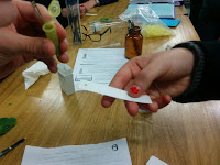
Separation of Chlorophyll Pigments by Paper

In my biology lesson we did a chromatography of chloroplast extract:

At first we added a spatula of anhydrous sodium sulphate into an Eppendorf tube.
Then we placed small pieces of spinach leaves into the tube and 5 drops of solvent A (2 parts ethyl ethanoate and 3 parts propanone) and stired al of it a metal probe:

Now we placed a dot of the solvent on a TLC plate with 5 mm space from the bottom edge:

After this we used a dropper pipette to place solvent B(5:3:2 mixture of cyclohexane, ethyl ethanoane and propanone) into a new vial (5mm hight of solution).
Finally we put the TLC plate into the vial and waited for a few minutes, so that the result was as shown here: